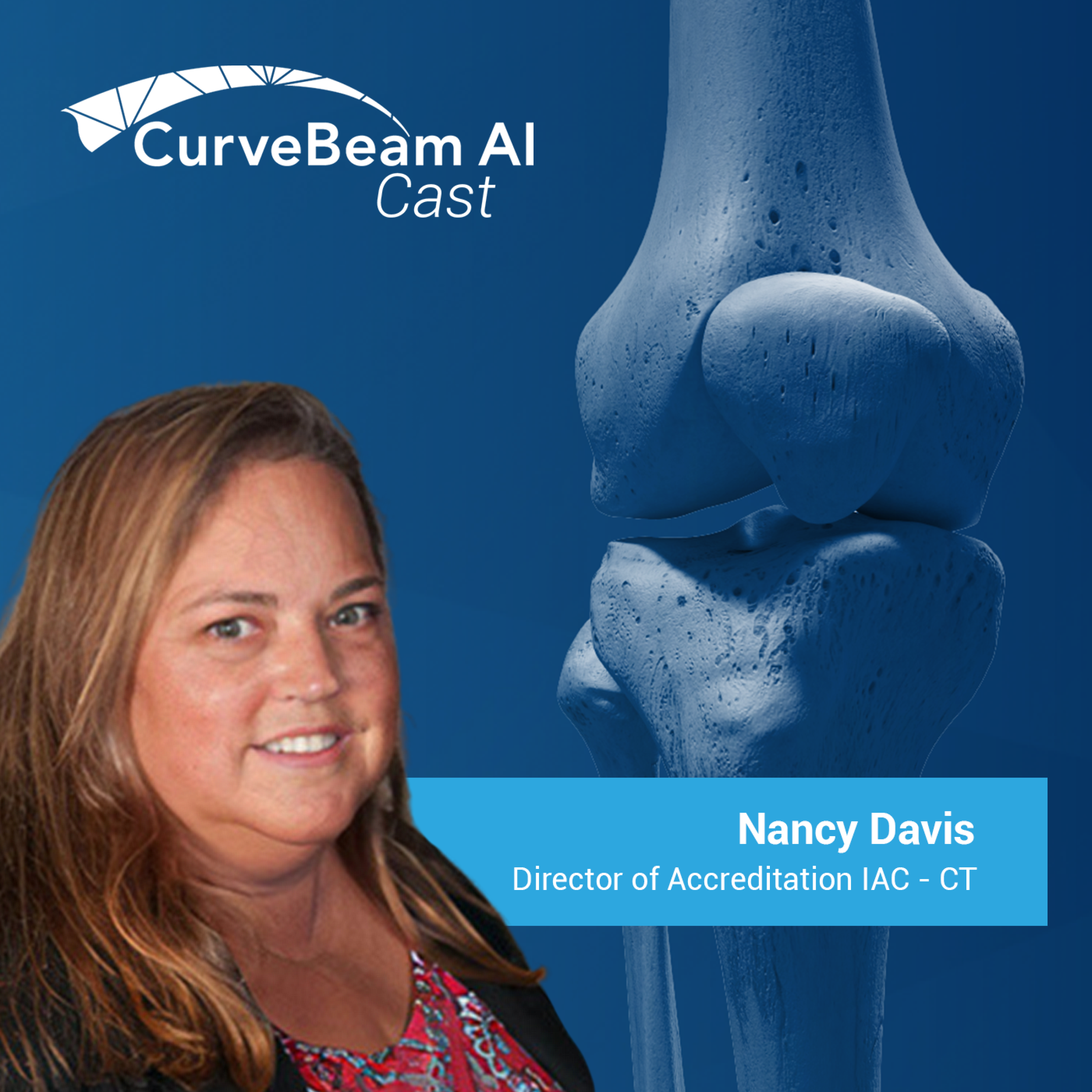
Biz and Tech Podcasts > Technology > CurveBeam AI Cast
Welcome to CurveBeam AI Cast, where we talk with doctors and experts in the field discussing innovations and insights into orthopedic imaging.
Last Episode Date: 14 August 2024
Total Episodes: 47
The integration of cone beam CT imaging in research labs is revolutionizing orthopedic investigations. Dr. Zong-Ming Li, PhD, David Jordan, and Trevour Greene from the University of Arizona discuss the transformative impact of the InReach CT system from CurveBeam AI on their work. The Hand Research Laboratory at the University of Arizona focuses on solving orthopedic-related clinical problems, including carpal tunnel syndrome and arthritis. The lab is equipped with various biomechanics tools, motion analysis systems, and two ultrasound machines, providing comprehensive facilities for their research.The introduction of the InReach CT system has significantly streamlined their workflow, allowing for immediate scanning without the constraints of clinical CT schedules. This flexibility enables the lab to conduct detailed biomechanical experiments, scanning both cadaveric and, eventually, human subjects with high resolution and low radiation dose. The convenience and ease of use of the InReach system have accelerated their research, allowing for quick quality control and immediate re-scanning if necessary.This episode highlights:The unique capabilities and comprehensive equipment of the Hand Research Laboratory.The benefits of the InReach CT system, including its low maintenance, flexibility, and high-resolution imaging.The application of their research findings to clinical settings, potentially improving diagnostic accuracy for joint space analysis.Dr. Li, Jordan, and Greene have established a dynamic research environment at the University of Arizona, leveraging advanced imaging technologies to further orthopedic research. Their work with the InReach system from CurveBeam AI exemplifies the transformative impact of innovative medical imaging tools on scientific investigations and clinical applications.
Rapid advancements in surgical planning techniques are propelling the field of orthopedic technology into a transformative phase. João Pedro Ribeiro, the Chairman and CEO of PeekMed, is at the forefront of this change. His story, from a biomedical engineer to an industry leader, highlights the impact of innovation in healthcare. As we enter an era where technology and medicine intersect more than ever, the stakes for improving patient outcomes have never been higher.So how does technology streamline surgical planning and enhance orthopedic care? This is the core question addressed in the latest episode of 'CurveBeam AI Cast'. Host Vinti Singh, Marketing Director at CurveBeam AI, welcomes João Pedro Ribeiro to discuss the evolution of orthopedic surgery with the aid of cutting-edge software platforms like PeekMed.This episode highlights:The origin and mission of PeekMed, emphasizing its role in pre-surgical planning.Insights on how PeekMed’s technology caters to the needs of both surgeons and the orthopedic industry, enhancing efficiency and precision.The future of orthopedics, where integration of comprehensive patient data could revolutionize treatment approaches.João Pedro Ribeiro, with his background in biomedical engineering and medical informatics, has established himself as an innovator in orthopedic technology. His leadership at PeekMed reflects his commitment to improving surgical outcomes through advanced planning tools, contributing significantly to the field of orthopedic surgery.
In the latest CurveBeam AI Cast episode, recorded at the AOFAS Annual Meeting 2023, host Dr. Lew Schon, MD, Director of Innovation at Mercy Medical Center and Professor at John Hopkins University and NYU Langone, engages in a riveting conversation with Dr. Marie-Aude Munoz, MD, a distinguished Orthopedic Foot & Ankle Surgeon from Montpellier, France. Together, they explore Dr. Munoz's trailblazing journey into opening a private practice to offer weight bearing CT imaging services to her patients.Discussion Highlights:• Dr. Munoz's transition from traditional orthopedics to establishing her own specialized medical center for foot and ankle pathology.• Her decision to be one of the first in France to offer weight-bearing CT technology imaging• An in-depth examination of how this imaging technology not only augments surgical planning but also enriches patient comprehension and satisfaction regarding their pathologiesDr. Marie-Aude Munoz, MD, is a pioneer in orthopedic surgery, specializing in foot and ankle pathologies. Her academic journey commenced in Paris, followed by specialization in Montpellier. With over a decade of experience in private practice, Dr. Munoz's visionary approach led to the creating of a specialized medical center for foot and ankle pathology, setting a notable precedent in the orthopedic community in France. Embracing the potential of weight-bearing CT technology, she continues to advance the standards of orthopedic healthcare.
The introduction of weight-bearing CT scans has revolutionized how surgeons assess and treat various orthopedic conditions. Research indicates this technology can provide clinicians with in-depth, real-time insights, allowing for better patient outcomes and more accurate diagnoses.How is weight bearing CT technology influencing the approach of orthopedic surgeons, and how are patients benefitting from it?Welcome to the CurveBeam AI Cast, where the future of orthopedics is at the forefront. In this episode, hosted by Dr. Lew Schon, a renowned Orthopedic Surgeon and Director of Innovation at Mercy Medical Center and Professor at John Hopkins University and NYU Langone, examines the impact of weight bearing CT imaging in a typical orthopedic clinic with his guest Dr. Blake Moore, an experienced Orthopedic Surgeon from Virginia Beach, who shares his journey with this innovative technology.Key conversation points include:• The evolution and early adoption of weight bearing CT scans• The physiological benefits of assessing limbs under actual weight bearing conditions• How the imaging precision of weight bearing CT scans influences surgical decision-makingDr. Blake Moore, originally from Albany, New York, completed his medical studies at Albany Medical College before undertaking his residency at Geisinger Medical Center in Pennsylvania. Following a fellowship with Paul Giuliano at Penn State, Dr. Moore has spent almost a decade in Virginia, using weight bearing CT scans in his practice and advocating for its broader implementation. His commitment to advancing orthopedics through technology and research is evident in his work and collaborative efforts with fellow professionals.
The benefits of WBCT (Weight Bearing Computed Tomography) usher in a new era of orthopedic diagnostics, illuminating previously obscure aspects of hindfoot alignment. The shift from 2D X-ray technology to 3D imaging has been profound, offering a more intricate understanding of bone structures and alignments, which is critical for effective treatment plans. Numerous research articles point to the reliability of different approaches in conventional X-ray and cone beam CT, showcasing the reliability of WBCT in hindfoot alignment assessment. As the dialogue around these advancements intensifies, the overarching query resonates within the medical community: How does this technological leap influence the accuracy and efficacy of orthopedic diagnostics and treatment?In this second of a two-part CurveBeam AI Cast series hosted by Dr. Lew Schon, MD, a renowned Orthopedic Surgeon and Director of Innovation at Mercy Medical Center and Professor at John Hopkins University and NYU Langone, continues his conversation with Dr. Francois Lintz, MD, an esteemed Orthopedic Foot & Ankle Surgeon and past President of the International WBCT Society. In this second conversation, Dr. Schon and Dr. Lintz look deeper into the groundbreaking advancements of 3D imaging and the benefits of WBCT.Highlights from Dr. Schon and Lintz’s second conversation include:• Dr. Lintz’s pursuit of expanding 3D biometric principles beyond the foot and ankle to encompass the entire lower limb, alongside standardizing these principles through an initiative called the “scanner's endeavor”• The introduction of a visualization tool known as “distance mapping” enabling a more nuanced analysis of joint space width, aiding in the differentiation between ligament laxity and early arthritis• The innovative exploration into bone density as a “fourth dimension” in orthopedic imaging, opening doors to more precise diagnosis and treatment planning
In recent years, the orthopedics field has been gravitating towards using 3D imaging technologies to foster a deeper understanding of bone structures and alignments. A notable driver of this shift is the application of Weight bearing Computed Tomography (WBCT), which has emerged as a game-changing tool for evaluating hindfoot alignment.What might the integration of weight bearing CT signify for the future of orthopedic diagnostics and treatment? How might this advancement in imaging technology alter the conventional practices in orthopedics?In the latest episode of CurveBeam AI Cast hosted by Dr. Lew Schon, MD, a renowned Orthopedic Surgeon, Director of Innovation at Mercy Medical Center and Professor at John Hopkins University and NYU Langone, a conversation unfolds with Dr. Francois Lintz, MD, FEBOT, an esteemed Orthopedic Foot & Ankle Surgeon and outgoing President of the International WBCT Society. The discussion, being the first of a two-part series, delves into the journey of Dr. Lintz in the realm of weight bearing CT and how this technology stands as a milestone in orthopedic imaging.Highlights from Dr. Schon and Lintz’s first conversation include:· The evolution of Dr. Lintz’s interest and work in weight-bearing CT from a project on hindfoot alignment in Bristol, UK, to the development of a 3D biometric system· Exploring the pivotal role of weight bearing 3D imaging technologies in advancing orthopedic practices, highlighting its significance in understanding and measuring complex bone alignments· Discussing the challenges and prospects in integrating weight-bearing CT technology into the mainstream orthopedic diagnostics and treatment protocols“This is a technology for everybody and that it comes in place of x-rays and CT, not just CT” Lintz said. “Weight bearing CT is in fact cone beam CT and it's a 3D x-ray.” WBCT is poised to become an integral part of doctors daily practice, eventually surpassing traditional X-rays, in Dr. Lintz’ opinion.Dr. Lintz is the outgoing president of the International WBCT Society. His educational journey and relentless pursuit of innovation have positioned him as a revered figure in the orthopedic community. His conversation with Dr. Schon sheds light on the transformative potential of WBCT in orthopedics, laying a path for further exploration and adoption of this groundbreaking technology.
Axiometrics may be new but it’s set on revolutionizing orthopedics forever. Every year, approximately two million Americans suffer from acute ankle sprains, and bone and joint injuries in the lower limbs. While most of these injuries heal with time, there is a subset of patients who experience chronic ankle instability, enduring persistent pain and limited mobility. Unfortunately, the underlying causes of this condition remain poorly understood. However, the tides may be turning, thanks to the perseverance of a determined doctor and advancements in imaging technology.Orthopedic surgeries play a crucial role in restoring mobility and alleviating pain for countless individuals. However, the complexity of these procedures and the potential for iatrogenic injuries necessitate constant innovation and improvement. By embracing new concepts, like axiometrics, which involve measuring the functional relationships between the hip, knee, and ankle joints, there is promise for identifying and addressing the underlying causes of joint deformities, ultimately leading to improved patient outcomes and a reduction in long-term complications. Dr. Andy Goldberg, Foot and Ankle Orthopedic Surgeon, developed axiometrics with CurveBeam AI to bring the axial plane back to the forefront in medical imaging. In a short documentary about CurveBeam AI‘s axiometrics, he explained how watching a loved one face chronic pain and deformity pushed his advocacy for revolutionary CT imaging of the lower extremities. Several doctors who work in the field of orthopedics have seen these joint injuries first-hand, how they develop, and what they believe is revolutionizing the field of orthopedics.Drs. Goldberg, San Giovanni, and Stronach’s Thoughts“So my interest in this area stemmed really back more than 20 years when my grandmother broke her hip and went into hospital, and she had her hip fixed with something called a dynamic hip screw. And it was done by a junior doctor, and I’m sure he’s well-meaning, but he rotated her femur slightly inwards and fixed it with a pin and plates. I noticed that her knee was facing inwards and her foot was facing inwards, which it wasn’t before. Over, I would say, the next year, she started to develop what’s called a valgus knee, so the knee started twisting outwards. And her foot started to collapse inwards, and she had what’s known as a planovalgus foot. This seemed to me to be an iatrogenic injury, meaning it was caused by the surgery that she’d had. And so I, again, went around trying to persuade doctors that this was an important area. Maybe we could look at twisting it back. And they all looked at me, with abhorrence that I could either question the surgery. They couldn’t demonstrate that it was rotated. On a plain x-ray, you can’t see that axial malrotation. Being in her late 80s, early 90s, no one wanted to operate to correct the problem. And over the next five years, she developed more and more deformity, more and more pain, more and more arthritis. For me, very disheartening that this was the experience that a patient would go through. And it stemmed my interest in a new type of surgery, which was a new type of surgery. I was very interested in this notion of axial rotation and the notion of axiometrics.”-Dr. Andy Goldberg, Foot and Ankle Orthopedic Surgeon“So we may see things downstream. It’s not uncommon that we’ll see six to nine months after a knee replacement in somebody that had, let’s say, a varus osteoarthritis of the knee, which might be the more common if it’s osteoarthritis, that will then start to have posterior tibial tendon dysfunction. Six, nine months later, start getting soreness. And they may have pre-existingly [sic] had some flat foot deformity, but asymptomatic, that has now become symptomatic.”-Dr. Thomas San Giovanni, MD, Foot and Ankle Orthopedic Surgeon“In general, we have seen increasing subspecialization of orthopedic surgeons where our focus has become more narrow. We see some surgeons that focus only on the hip, or only on the knee. Foot and ankle specialists really focus only on that area. And so we do see increasing subspecialization in our field. It really means we’ve got to be very good about communication because it can have significant impact on your area that you’re treating, such as how the ankle can affect the hip, and these other areas are all connected to one another.”-Dr. Benjamin M. Stronach, Hip and Knee Orthopedic Surgeon
Imaging and diagnostics are important facets in healthcare and nearly every area of medicine relies on this technology to diagnose or monitor conditions. In orthopedics, it is an especially vital tool, as it allows surgeons to discover and perform successful procedures for their patients. Currently, improved AI imaging technology is helping to support orthopedic surgeons in identifying and better detecting concerns and conditions. CurveBeam AI is a leader in developing equipment to assist orthopedic surgeons in this process and will help change the future of AI imaging technology and orthopedics.In what way is AI imaging technology being used in orthopedics and how will it revolutionize surgical procedures?To explore the topic in the “CurveBeam AI” podcast, Dr. Lew C. Schon, orthopedic surgeon and professor, interviewed Stuti Singh, the company’s Director of Imaging and Diagnostics. The two discussed the general expectations of imaging and diagnostics, AI imaging technology, and where AI stands to be a gamechanger in the imaging field.Dr. Schon and Singh also talked more on the subject in regards to … 1. The challenges pertaining to metal hardware and how it affects imaging but also assists CurveBeam AI in improving their technology2. How 2D images are converted to 3D images for higher quality and better orthopedic estimations3. How data and troubleshooting play a big factor in CurveBeam AI’s algorithm“We will have customers send us examples of images they’re struggling with, of anatomy they want to see, but it’s too blurry… and we actually use those images that we get in our development. So, we will test newer versions of the algorithm with that clinical data, with their permission, of course. We actually even have some customers who have, in the validation of this product, to replace their multi-slice CT. They have scanned a patient with metal hardware in multi-slice CT and in cone-beam CT and have shared the difference with us so we can see kind of what the more ideal image would look like on multi-slice CT and having that kind of ground truth is really useful as well. So, we work really closely with our customers and it's really valuable, I think, going both ways because we are hopefully giving them the better and better product, but it’s really their feedback and their data that helps us get there,” said Singh.Dr. Lew C. Schon is a board-certified orthopedic surgeon and is the Director of Orthopedic Innovation Institute of Foot and Ankle Reconstruction at Mercy Medical Center in Baltimore. He is also a professor of orthopedic surgery at NYU Langone and Johns Hopkins University, respectively. Stuti Singh is the Director of Imaging and Diagnostics at CurveBeam AI. She has held that role for over a year now and has been with the company for more than eight years. Prior to that she worked as an engineer for Boeing. Singh is also an alum of Georgia Institute of Technology.
The foot and ankle are complex structures comprised of 26 small bones and 29 muscles. With so many structures and an incredible amount of variation in how deformities can present, surgical corrections aren’t so simple. For example, there are more than 100 operations that can be performed for hallux valgus deformities, commonly known as bunions. 3D planning helps to address the variations and nuances of foot and ankle surgeries to assist in obtaining optical post-surgical outcomes.How is one company, RedPoint Medical 3D, making 3D planning more accessible to orthopedic surgeons?On this episode of CurveBeam AI, host Lew Schon, MD, Director of Orthopedic Innovation at the Institute of Foot and Ankle Reconstruction at Mercy Medical Center, speaks with Dr. Adam Perler, DPM, Co-Founder & Chief Innovations Officer at RedPoint Medical 3D and Surgeon at Alexander Orthopaedic Associates, to discuss the RedPoint Medical 3D has created a vendor-agnostic, streamlined process for 3D modeling, custom surgical planning, and 3D printed cut guides.Dr. Schon and Dr. Perler also discussed…1. The role that 3D printing plays in helping surgeons visualize their surgical approaches2. How older and more experienced surgeons have reacted to the redpointing philosophy3. What the process of redpointing looks like from receiving a 3D CT scan to the point of the surgical procedureDr. Perler explained how RedPoint Medical 3D was named. “Redpointing is where a professional climber will write down all the details of the route, where they’re going to place their hands, where they’re going to place their feet. And then they hand that plan to a judge and they don’t actually touch the rock face until they hand the plan over. Then they go about their climb. If they do it perfectly, that’s called redpointing. So, to me, this was a vision of what I wanted to be able to help surgeons do with their surgeries and I would love to get to the point where redpointing becomes a very grammatical term for us all to describe, ‘Hey, I’ve got this big surgery coming up’ and another surgeon says, ‘Hey, are you going to redpoint that?’ meaning are you going to meticulously plan that and flawlessly execute that?”Dr. Adam Perler, Doctor of Podiatric Medicine (DPM), is an experienced foot and ankle surgeon passionate about new product development for surgeries. He is Co-Founder & Chief Innovations Officer at RedPoint Medical 3D and Surgeon at Alexander Orthopedic Associates. Dr. Perler earned his B.A. in Biology from Indiana University Bloomington and his DPM from Dr. William M. Scholl College of Podiatric Medicine at Rosalind Franklin University of Medicine. He is residency-trained at Palmetto General Hospital’s Surgical Residency and later pursued fellowship training from the Ilizarov Orthopedic Lower Extremity Reconstructive Fellowship and the AO Trauma Fellowship in Hannover, Germany.
Dr. Lew Schon, MD, makes his debut as guest host on CurveBeam AI Cast in an interview with his former fellow, Dr. Cesar de Cesar Netto, MD, PhD. The two reminisce about how Dr. De Netto was first introduced to WBCT imaging at Medstar Union Memorial Hospital in Baltimore, and how he is now a global leader in WBCT research and applications.The two discuss:Dr. de Netto's research into progressive collapsing foot disorder (PCFD) and how WBCT could guide treatment, for example fusion vs. joint preserving procedures.Whether Dr. de Netto thinks all foot & ankle studies will eventually need to be redone using WBCT (spoiler: yes).How Dr. de Netto's patients are specifically seeking him out to assess their foot & ankle pain with WBCT imaging.Click here to read about a research project in which Dr. de Netto's team combined WBCT imaging with coverage mapping and distance mapping to better understand bone positioning in patients with progressive collapsing foot deformity (PCFD).Dr. Cesar de Cesar Netto is an Assistant Professor at the University of Iowa, Carver College of Medicine, specifically in the Department of Orthopedics and Rehabilitation. Dr. de Cesar Netto has had four Clinical Fellowships, including at the University of Sao Paulo, the University of Alabama, the Hospital for Special Surgery in New York, and the Medstar Union Memorial Hospital in Baltimore. He specializes in Foot and Ankle Surgery Disorders and Imaging of the Foot and Ankle, with a focus on Flatfoot Deformity, Tomography and Achilles Tendinopathy.
Discover new partners and
collaboration opportunities —right in your inbox.
Get notified about new partnerships